Dr. Hiram Fischer Trindade, European Implantology Center EIC, Portugal.
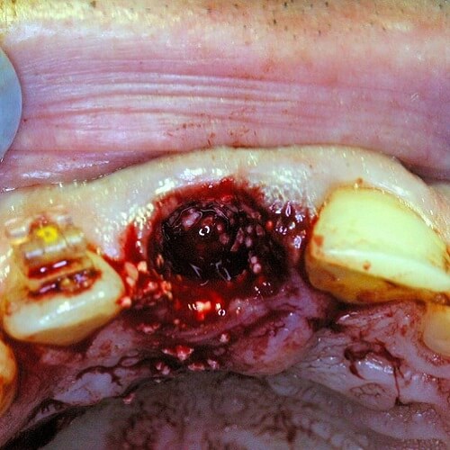
Bone Graft: 0,5g adboneTCP (0.1-0.5 mm).
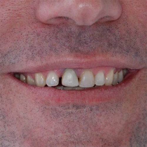
Patient: Male, 35 years old.
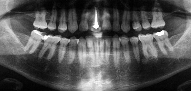
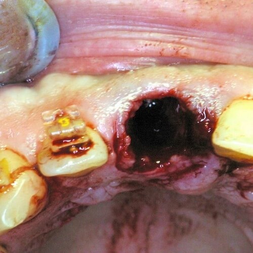
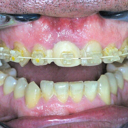
A 35-year-old male patient presented a severe root resorption.
Severe root resorption is very difficult to treat and often requires the extraction of teeth.
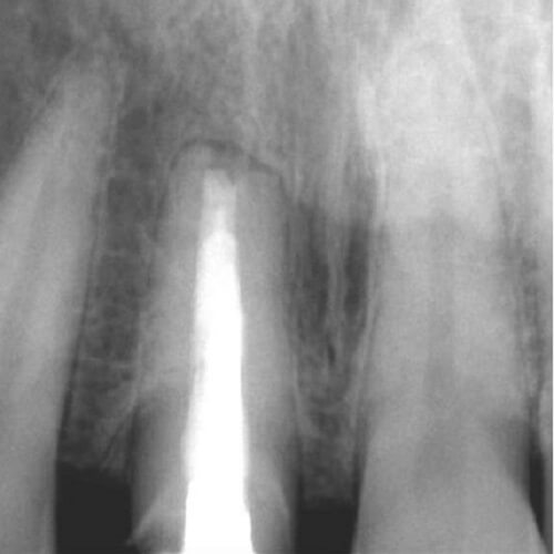
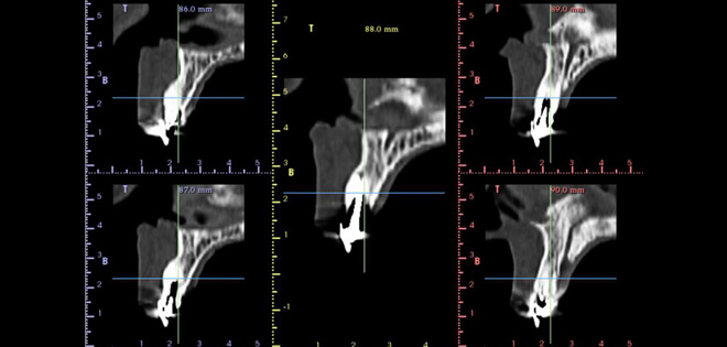
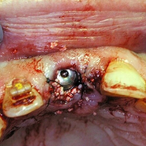
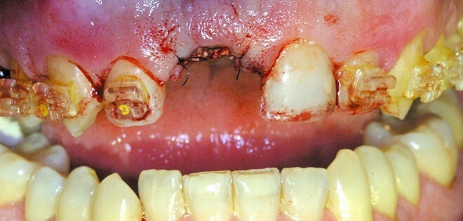
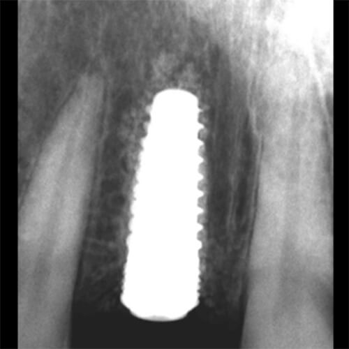
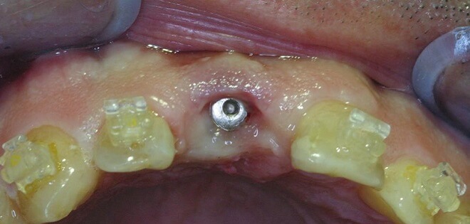
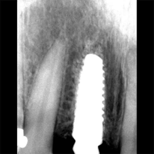
The surgical phase was initiated with the removal of UR1. It was used 0,5g of adbone®TCP, 0.1 – 0,5 mm granules, due to a severe bone loss.
The implant was placed at the time of grafting.
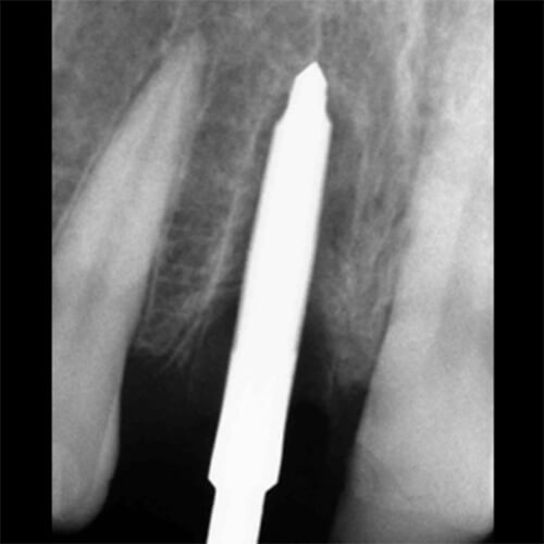
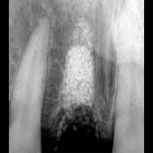
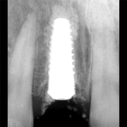
Radiographically, the bone density appeared to have improved in the UR1 place.