Dr. Reiser & Dr. Zinger, Tel-Aviv Sourasky medical center.
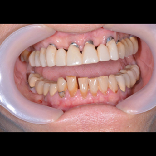
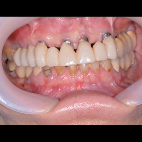
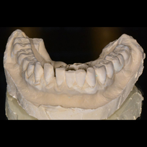
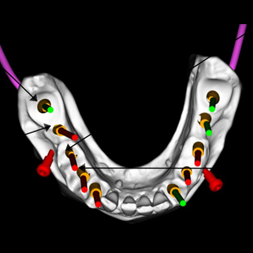
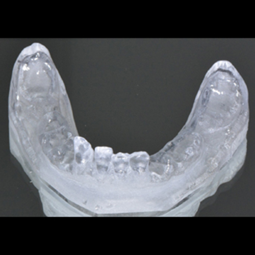
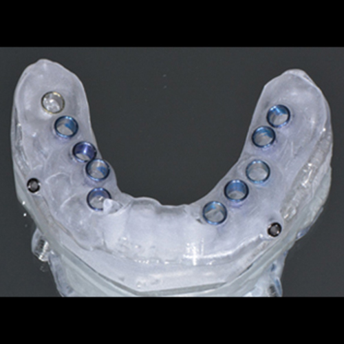
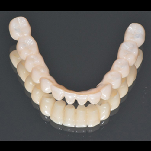
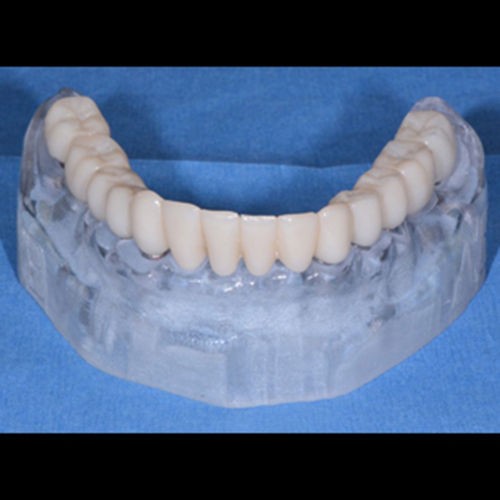
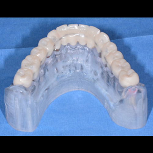
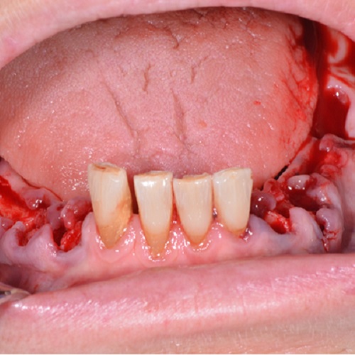
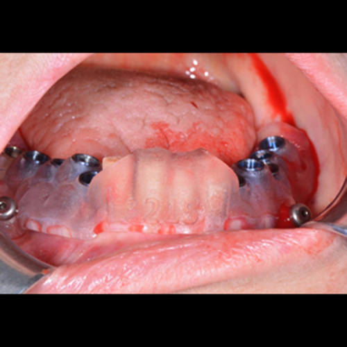
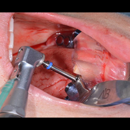
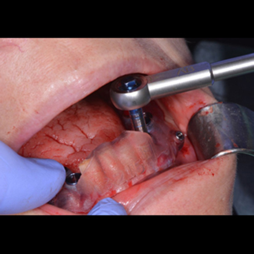
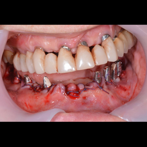
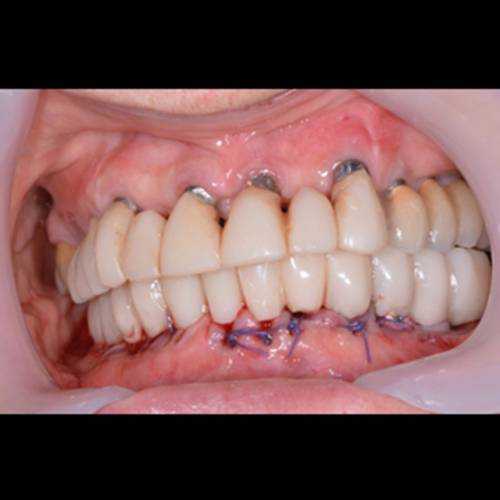
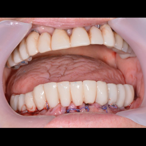
This patient requires extraction of all teeth in the lower jaw, immediate implant placement, and immediate loading with a CAD/CAM bridge prepared in advance on a digitally manufactured analog model.
This case presentation shows the digital process.
All the planning and production are provided by ABGuidedService, which gives a complete solution for each case.
The implants, planning, abutments, models and bridge are all from ABGuidedService.