Dr. Babich Semion.
There is a general misconception that Guided Implantology is only for complicated cases. However, there is great value in using this technology for even the “easy” cases.
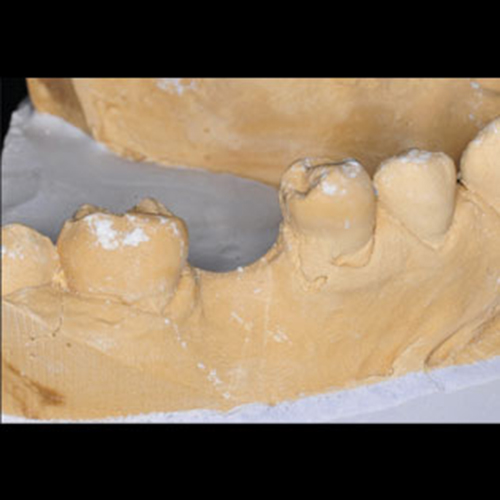
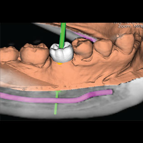
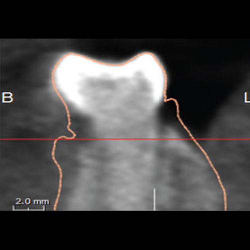
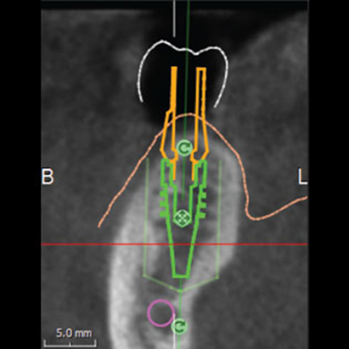
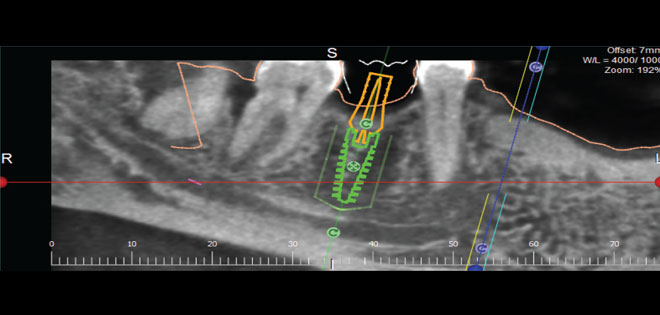
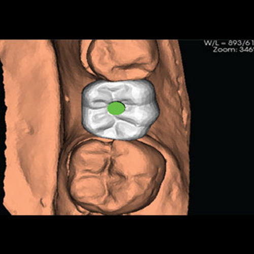
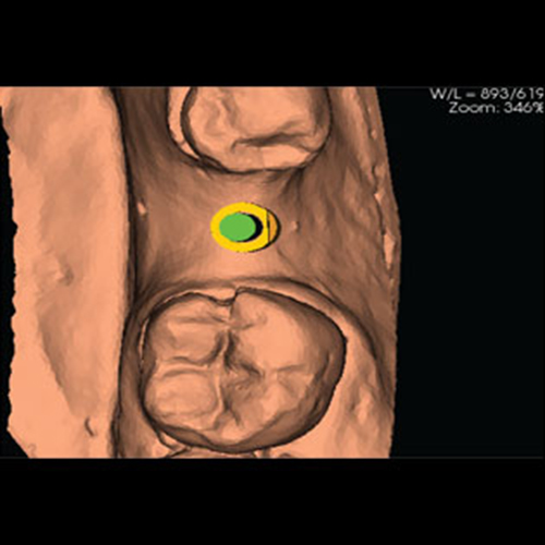
The 3D planning software allows us to relate to all the 5 directions: mesial, distal, buccal, lingual and depth. The implant can be placed in exactly the right position, and often with minimally invasive flapless surgery, with the use of a surgical guide. All the calculations are made before the surgery, and the use of drills with stoppers for the required depth, gives maximum safety.
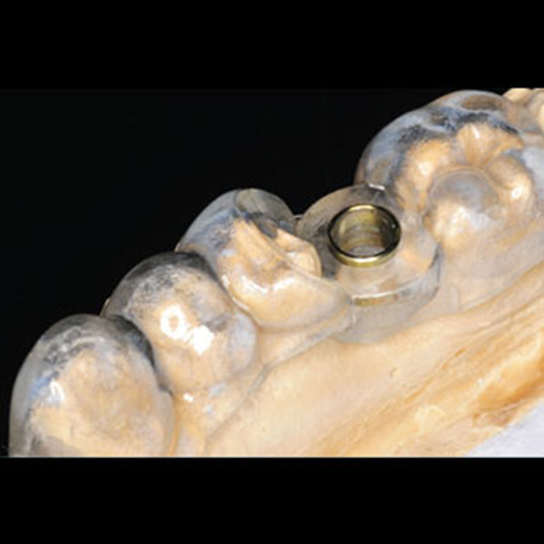
The implant is inserted through the guide with depth controlled implant mounts. The same procedure is used for multiple implants, for accurate and predictable results.